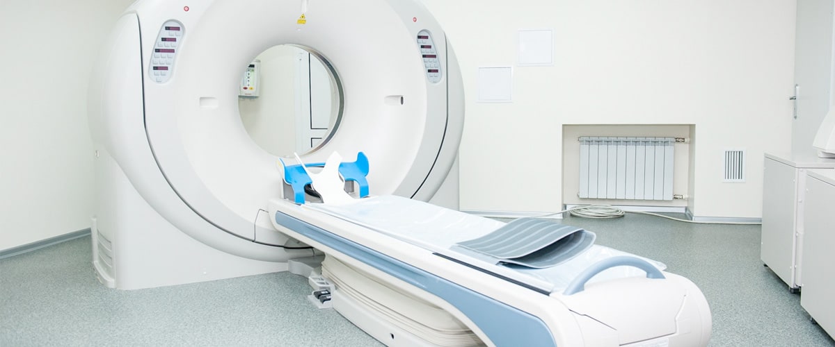
Computerised Tomography (CT) or scanner is a diagnostic medical technique which uses X-rays. From its appearance in the 1970s there have been great advances in this technique with the arrival of the helical scanner and multi-detector system, reducing the run-time and radiation of the scans whilst increasing the quality of the images.
It distinguishes between the different tissues by their resistance to the passing of radiation and can be applied at all levels of our anatomy: nervous system, thorax abdomen, skeleton, cardio-vascular system, etc.
Our equipment reduces the dose of radiation received by 70% and allows all the above mentioned studies to be carried out, as well as dental studies of the highest quality with minimum radiation.
Over recent years with technological advances a significant reduction in the dose of radiation received has been achieved and this allows it to be used not only as a diagnostic technique but also one for early detection of disease. For example, minimal radiation chest scans are now available which can detect early signs of lung cancer, a tumour that previosuly had a high mortality rate due to its late-stage diagnosis.
Also, by means of a specific program, we are able to carry out a virtual colonoscopy. This is when we see our colon without introducing a tube (it is recommended that colonoscopies are carried out regularly from the age of 50) and allows for the early detection and monitoring of colon cancer.


